Medical imaging allows doctors to examine structures inside the body without surgery, but different types of scans work in different ways. CAT scans, also known as CT or computed tomography scans, use X-rays and computer processing to produce detailed cross-sectional images. They are commonly used to investigate injuries, internal bleeding, fractures, lung conditions and abnormalities affecting organs and blood vessels.
An MRI scan, or magnetic resonance imaging examination, uses a strong magnetic field and radio waves rather than X-rays to create detailed images of structures inside the body. MRI is particularly useful for examining soft tissues, including the brain, spinal cord, muscles, ligaments, tendons and joints. The choice between CT and MRI depends on the part of the body being investigated and the information the doctor needs.
How CAT Scans Work
During a CT examination, the patient lies on a table that moves through a large, ring-shaped scanner. An X-ray source rotates around the body and captures information from multiple angles. A computer then processes this information to create cross-sectional images.
One major advantage of CT is speed. Scans can often be completed quickly, making CT particularly useful in emergency situations. For example, doctors may request a CT examination following a serious accident to rapidly investigate internal injuries or bleeding.
Contrast material may sometimes be used to make particular tissues, organs or blood vessels easier to see. Depending on the examination, contrast may be given orally or through an intravenous injection.
How MRI Works
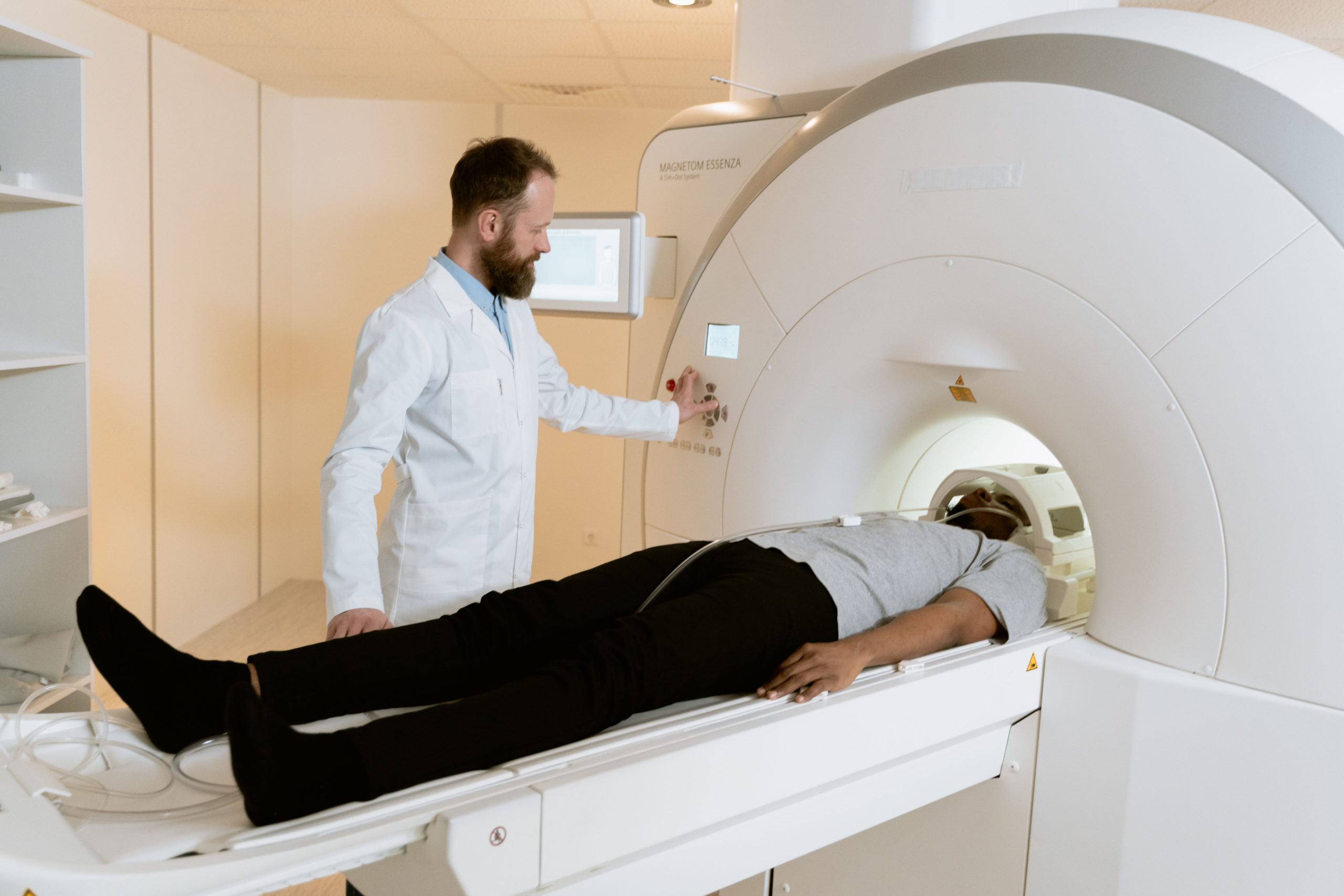
An MRI machine uses powerful magnets and radiofrequency energy to generate detailed images. Patients usually lie inside a tube-shaped scanner while the examination is performed.
MRI examinations generally take longer than CT scans and require patients to remain still for an extended period. The machine also produces loud knocking or tapping sounds during imaging, so patients are usually provided with hearing protection.
Because MRI does not use ionising radiation, it can be useful when detailed soft-tissue imaging is required. However, the strong magnetic field means patients must complete safety screening before entering the scanning room. Certain metallic implants, medical devices or metal fragments can affect whether an MRI is appropriate.
What Can Each Scan Detect?
CT provides excellent detail of bones, lungs and many internal organs. It is frequently used to investigate fractures, chest conditions, kidney stones, internal injuries and some cancers. It can also be used to guide certain medical procedures.
MRI generally provides greater soft-tissue contrast. Doctors may request it when examining the brain, spinal cord, joints, muscles or other soft tissues. It can help investigate conditions involving ligaments, nerves and neurological structures.
There are situations where either examination could provide useful information. In others, one technique may be clearly better suited to the medical question being investigated.
Which Scan Should You Have?
Patients generally do not need to decide between CT and MRI themselves. A GP or specialist will recommend the appropriate examination based on symptoms, medical history and the area requiring investigation.
Cost, availability, urgency and individual safety considerations can also influence which examination is appropriate. Patients who are pregnant, have implanted medical devices or have previously experienced reactions to contrast material should discuss this with their doctor and imaging provider.
Both CT and MRI are valuable diagnostic technologies, but they produce images using very different methods. Understanding these differences can help patients know what to expect while their healthcare professional determines which examination is most appropriate for their individual medical needs.